Category:Brain MRI glioma case
Jump to navigation
Jump to search
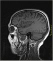
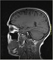
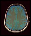
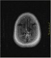
English: Brain MRI & Spectroscopy of 3cm low grade glioma located at left insula 52f. MRS shows increased Choline, decrease NAA, Cho NAA ratio 3.15, no lactate and lipid.


False-color Brain MRI
| This image set can be scrolled interactively with the mouse (wheel or dragging). This is done by using the template Imagestack. |
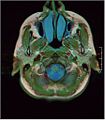
Trichromatic, False color MRI
- T1 in Red channel,
- PD in Green channel,
- T2 in Blue channel.
Key to colors:
- Cyan / Blue : Water, Cerebrospinal fluid, Eyes, Edema
- Green: Gray matter
- Light Green: Parotis, Salivary glands
- Dark Green: Muscles
- Brown / red : White matter
- Red: Large arteries
- Dark red: Veins
- Whitish yellow: Fat
- Black: Air, signal void
Media in category "Brain MRI glioma case"
The following 101 files are in this category, out of 101 total.
-
 Brain MRI glioma 00.jpg 448 × 512; 25 KB
-
 Brain MRI glioma 01.jpg 448 × 512; 26 KB
-
 Brain MRI glioma 02.jpg 448 × 512; 27 KB
-
 Brain MRI glioma 021.jpg 448 × 512; 32 KB
-
 Brain MRI glioma 022.jpg 448 × 512; 34 KB
-
 Brain MRI glioma 023.jpg 448 × 512; 36 KB
-
 Brain MRI glioma 024.jpg 448 × 512; 36 KB
-
 Brain MRI glioma 025.jpg 448 × 512; 38 KB
-
 Brain MRI glioma 026.jpg 448 × 512; 38 KB
-
 Brain MRI glioma 027.jpg 448 × 512; 40 KB
-
 Brain MRI glioma 028.jpg 448 × 512; 40 KB
-
 Brain MRI glioma 029.jpg 448 × 512; 41 KB
-
 Brain MRI glioma 03.jpg 448 × 512; 27 KB
-
 Brain MRI glioma 030.jpg 448 × 512; 41 KB
-
 Brain MRI glioma 031.jpg 448 × 512; 42 KB
-
 Brain MRI glioma 032.jpg 448 × 512; 41 KB
-
 Brain MRI glioma 033.jpg 448 × 512; 40 KB
-
 Brain MRI glioma 034.jpg 448 × 512; 39 KB
-
 Brain MRI glioma 04.jpg 448 × 512; 26 KB
-
 Brain MRI glioma 040.jpg 448 × 512; 34 KB
-
 Brain MRI glioma 043.jpg 448 × 512; 40 KB
-
 Brain MRI glioma 044.jpg 448 × 512; 41 KB
-
 Brain MRI glioma 045.jpg 448 × 512; 41 KB
-
 Brain MRI glioma 046.jpg 448 × 512; 43 KB
-
 Brain MRI glioma 047.jpg 448 × 512; 42 KB
-
 Brain MRI glioma 048.jpg 448 × 512; 43 KB
-
 Brain MRI glioma 05.jpg 448 × 512; 26 KB
-
 Brain MRI glioma 051.jpg 448 × 512; 44 KB
-
 Brain MRI glioma 06.jpg 448 × 512; 27 KB
-
 Brain MRI glioma 060.jpg 448 × 512; 37 KB
-
 Brain MRI glioma 061.jpg 448 × 512; 36 KB
-
 Brain MRI glioma 062.jpg 448 × 512; 35 KB
-
 Brain MRI glioma 063.jpg 294 × 384; 32 KB
-
 Brain MRI glioma 064.jpg 294 × 384; 32 KB
-
 Brain MRI glioma 065.jpg 294 × 384; 33 KB
-
 Brain MRI glioma 066.jpg 294 × 384; 33 KB
-
 Brain MRI glioma 068.jpg 294 × 384; 33 KB
-
 Brain MRI glioma 069.jpg 294 × 384; 32 KB
-
 Brain MRI glioma 07.jpg 448 × 512; 28 KB
-
 Brain MRI glioma 070.jpg 294 × 384; 32 KB
-
 Brain MRI glioma 071.jpg 294 × 384; 32 KB
-
 Brain MRI glioma 073.jpg 294 × 384; 32 KB
-
 Brain MRI glioma 074.jpg 294 × 384; 32 KB
-
 Brain MRI glioma 075.jpg 294 × 384; 31 KB
-
 Brain MRI glioma 076.jpg 294 × 384; 31 KB
-
 Brain MRI glioma 077.jpg 294 × 384; 30 KB
-
 Brain MRI glioma 079.jpg 294 × 384; 30 KB
-
 Brain MRI glioma 08.jpg 448 × 512; 29 KB
-
 Brain MRI glioma 080.jpg 294 × 384; 29 KB
-
 Brain MRI glioma 081.jpg 294 × 384; 29 KB
-
 Brain MRI glioma 082.jpg 294 × 384; 29 KB
-
 Brain MRI glioma 083.jpg 316 × 320; 17 KB
-
 Brain MRI glioma 084.jpg 316 × 320; 18 KB
-
 Brain MRI glioma 085.jpg 316 × 320; 19 KB
-
 Brain MRI glioma 086.jpg 316 × 320; 20 KB
-
 Brain MRI glioma 087.jpg 316 × 320; 21 KB
-
 Brain MRI glioma 088.jpg 316 × 320; 21 KB
-
 Brain MRI glioma 089.jpg 316 × 320; 22 KB
-
 Brain MRI glioma 09.jpg 448 × 512; 29 KB
-
 Brain MRI glioma 090.jpg 316 × 320; 22 KB
-
 Brain MRI glioma 091.jpg 316 × 320; 23 KB
-
 Brain MRI glioma 092.jpg 316 × 320; 24 KB
-
 Brain MRI glioma 093.jpg 316 × 320; 24 KB
-
 Brain MRI glioma 094.jpg 316 × 320; 23 KB
-
 Brain MRI glioma 095.jpg 316 × 320; 23 KB
-
 Brain MRI glioma 096.jpg 316 × 320; 22 KB
-
 Brain MRI glioma 097.jpg 316 × 320; 21 KB
-
 Brain MRI glioma 098.jpg 316 × 320; 21 KB
-
 Brain MRI glioma 099.jpg 316 × 320; 20 KB
-
 Brain MRI glioma 10.jpg 448 × 512; 30 KB
-
 Brain MRI glioma 101.jpg 316 × 320; 19 KB
-
 Brain MRI glioma 102.jpg 316 × 320; 17 KB
-
 Brain MRI glioma 103.jpg 448 × 512; 36 KB
-
 Brain MRI glioma 104.jpg 448 × 512; 37 KB
-
 Brain MRI glioma 105.jpg 448 × 512; 36 KB
-
 Brain MRI glioma 106.jpg 448 × 512; 37 KB
-
 Brain MRI glioma 107.jpg 448 × 512; 37 KB
-
 Brain MRI glioma 108.jpg 448 × 512; 36 KB
-
 Brain MRI glioma 109.jpg 448 × 512; 36 KB
-
 Brain MRI glioma 11.jpg 448 × 512; 31 KB
-
 Brain MRI glioma 110.jpg 448 × 512; 36 KB
-
 Brain MRI glioma 111.jpg 448 × 512; 37 KB
-
 Brain MRI glioma 112.jpg 448 × 512; 37 KB
-
 Brain MRI glioma 113.jpg 448 × 512; 38 KB
-
 Brain MRI glioma 114.jpg 448 × 512; 39 KB
-
 Brain MRI glioma 115.jpg 448 × 512; 39 KB
-
 Brain MRI glioma 116.jpg 448 × 512; 39 KB
-
 Brain MRI glioma 117.jpg 448 × 512; 41 KB
-
 Brain MRI glioma 118.jpg 448 × 512; 41 KB
-
 Brain MRI glioma 119.jpg 448 × 512; 42 KB
-
 Brain MRI glioma 12.jpg 448 × 512; 31 KB
-
 Brain MRI glioma 121.jpg 448 × 512; 40 KB
-
 Brain MRI glioma 13.jpg 448 × 512; 32 KB
-
 Brain MRI glioma 14.jpg 448 × 512; 34 KB
-
 Brain MRI glioma 15.jpg 448 × 512; 34 KB
-
 Brain MRI glioma 16.jpg 448 × 512; 35 KB
-
 Brain MRI glioma 17.jpg 448 × 512; 35 KB
-
 Brain MRI glioma 18.jpg 448 × 512; 34 KB
-
 Brain MRI glioma 19.jpg 448 × 512; 34 KB
-
 Brain MRI glioma 20.jpg 448 × 512; 34 KB
-
 MRS 120511.png 1,011 × 606; 15 KB


